Home

Straße Misty schwimmend stroke volume echo Beton Unterdrücker Schmiede

Management of Shock: Bedside Assessment of Cardiac Output EMRA

Comparison of pulse pressure variation versus echocardiography-derived stroke  volume variation for prediction of fluid responsiveness in mechanically  ventilated anesthetized dogs - Veterinary Anaesthesia and Analgesia
Comparison of pulse pressure variation versus echocardiography-derived stroke volume variation for prediction of fluid responsiveness in mechanically ventilated anesthetized dogs - Veterinary Anaesthesia and Analgesia

Normal Values of Cardiac Output and Stroke Volume According to Measurement  Technique, Age, Sex, and Ethnicity: Results of the World Alliance of  Societies of Echocardiography Study - Journal of the American Society
Normal Values of Cardiac Output and Stroke Volume According to Measurement Technique, Age, Sex, and Ethnicity: Results of the World Alliance of Societies of Echocardiography Study - Journal of the American Society

Calculation of the stroke volume (SV) and cardiac output (CO) by... |  Download Scientific Diagram
Calculation of the stroke volume (SV) and cardiac output (CO) by... | Download Scientific Diagram

Cardiac Output by Echo - INTENSIVE

How To Calculate Stroke Volume In Echocardiography (The EASY Way)

Accurate stroke volume (SV) estimation: SV = LVOT area × LVOT VTI. a... |  Download Scientific Diagram
Accurate stroke volume (SV) estimation: SV = LVOT area × LVOT VTI. a... | Download Scientific Diagram

3 Steps to Quantifying MR via Stroke Volume Method!

3 Steps to Quantifying MR via Stroke Volume Method!

A, Normal LVOT VTI (VTI TSVI, 19.09 cm), indicating a normal stroke... |  Download Scientific Diagram
A, Normal LVOT VTI (VTI TSVI, 19.09 cm), indicating a normal stroke... | Download Scientific Diagram

Cardiac Output
Cardiac Output

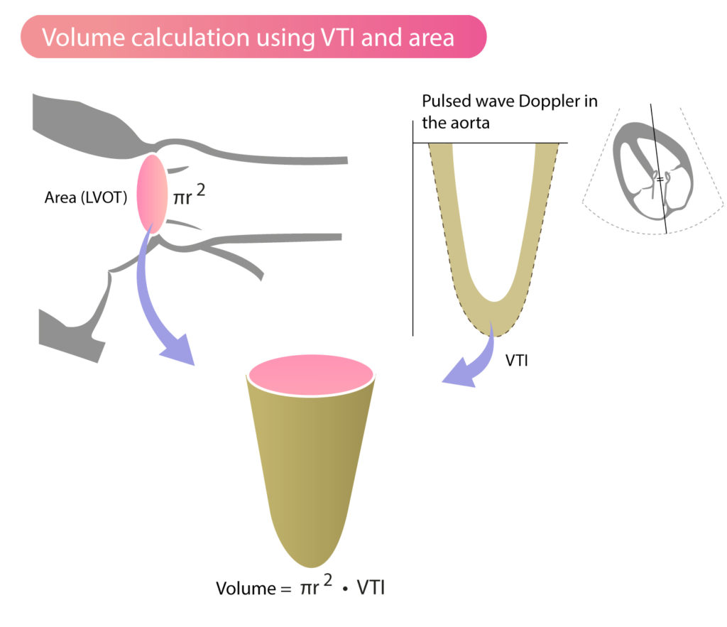
What is time velocity Integral ? (VTI ) | Dr.S.Venkatesan MD

Estimation of Stroke Volume and Aortic Valve Area in Patients with Aortic  Stenosis: A Comparison of Echocardiography versus Cardiovascular Magnetic  Resonance - Journal of the American Society of Echocardiography
Estimation of Stroke Volume and Aortic Valve Area in Patients with Aortic Stenosis: A Comparison of Echocardiography versus Cardiovascular Magnetic Resonance - Journal of the American Society of Echocardiography
3 Steps to Quantifying MR via Stroke Volume Method!

Hemodynamics | Thoracic Key

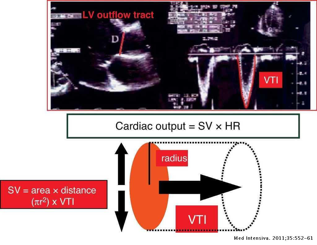
Estimating cardiac output. Utility in the clinical practice. Available  invasive and non-invasive monitoring | Medicina Intensiva
Estimating cardiac output. Utility in the clinical practice. Available invasive and non-invasive monitoring | Medicina Intensiva

Hemodynamics - Echocardiografie.nl

Stroke Volume & Cardiac Output Measurement on POCUS Accurately ... | GrepMed

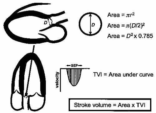
Stroke Volume, VTI (Velocity Time Integral) & Cardiac Output – ECG & ECHO

How To Calculate Stroke Volume In Echocardiography (The EASY Way)

LVOT Doppler for estimation of cardiac output | Deranged Physiology

Point of Care Echo: Stroke Volume Determination - YouTube

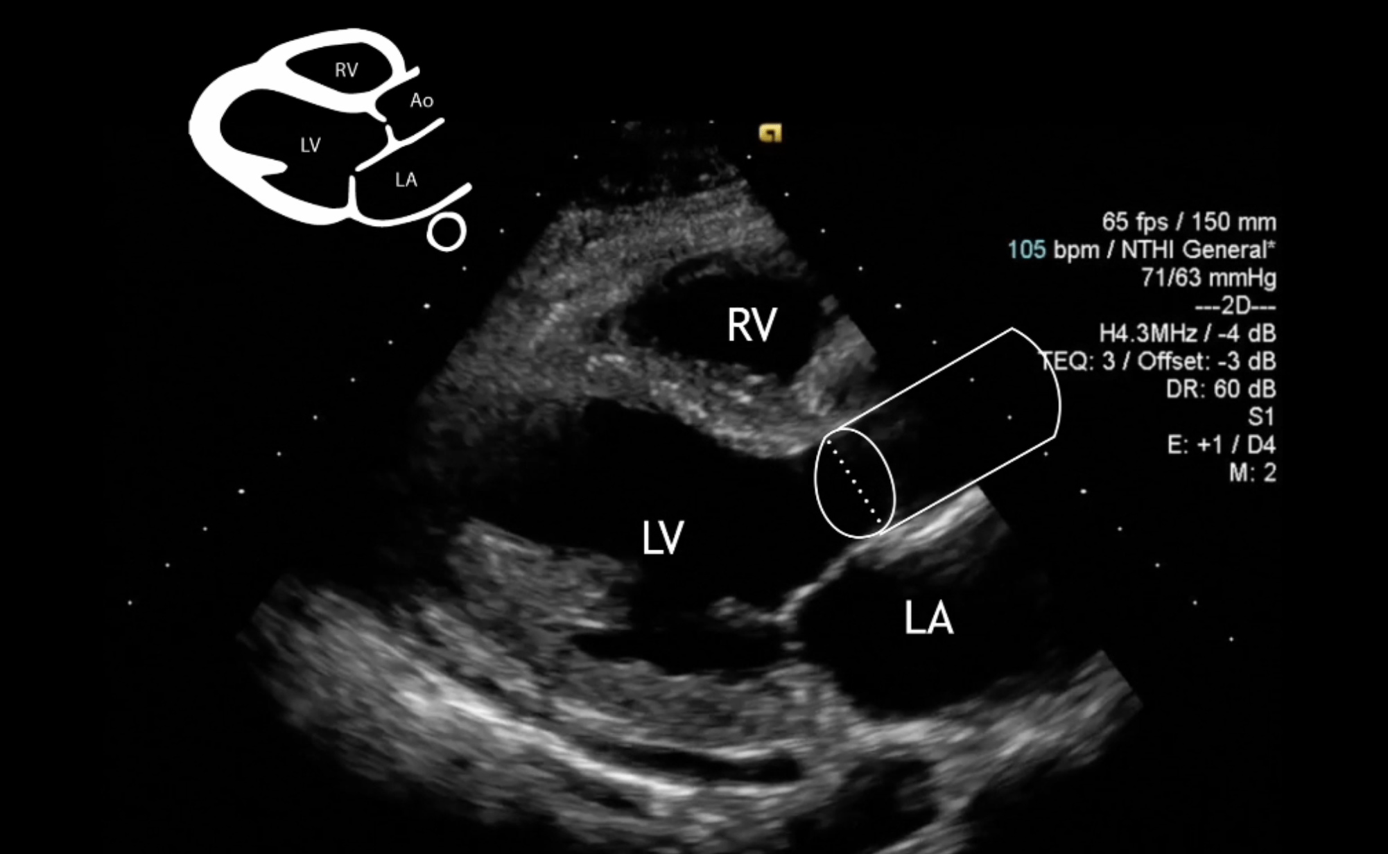
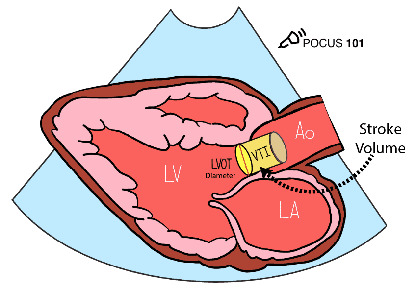
Measuring Cardiac Output with Echocardiography Made Easy - POCUS 101

Prognosis of Severe Low-Flow, Low-Gradient Aortic Stenosis by Stroke Volume  Index and Transvalvular Flow Rate | JACC: Cardiovascular Imaging
Prognosis of Severe Low-Flow, Low-Gradient Aortic Stenosis by Stroke Volume Index and Transvalvular Flow Rate | JACC: Cardiovascular Imaging

Fig. 18. (A) Stroke volume by Doppler (LVOT). (B) Stroke volume by Doppler  (mitral inflow). (C)… | Diagnostic medical sonography, Cardiac sonography,  Echocardiogram
Fig. 18. (A) Stroke volume by Doppler (LVOT). (B) Stroke volume by Doppler (mitral inflow). (C)… | Diagnostic medical sonography, Cardiac sonography, Echocardiogram
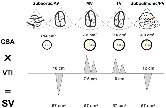
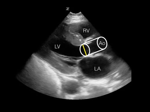
Right Ventricular Stroke Volume from Doppler

Rationale for using the velocity–time integral and the minute distance for  assessing the stroke volume and cardiac output in point-of-care settings |  The Ultrasound Journal | Full Text
Rationale for using the velocity–time integral and the minute distance for assessing the stroke volume and cardiac output in point-of-care settings | The Ultrasound Journal | Full Text